Isolation of Salmonella from raw beef and chicken used in fast food restaurants in Abuja, Nigeria
Salome Samuel Bawa, Jacob Kwada Paghi Kwaga, Mohammed Kabiru Lawan, Samuel Bitrus Bawa
Corresponding author: Salome Samuel Bawa, Department of Veterinary and Pest Control Services, Federal Ministry of Agriculture and Rural Development, No1 Kapital Street, Garki Area 11 Abuja, Nigeria 
Received: 12 Jun 2020 - Accepted: 18 Nov 2020 - Published: 25 Nov 2020
Domain: Public health
Keywords: Foodborne, Salmonella, fast-food, resistance, invA gene
©Salome Samuel Bawa et al. PAMJ-One Health (ISSN: 2707-2800). This is an Open Access article distributed under the terms of the Creative Commons Attribution International 4.0 License (https://creativecommons.org/licenses/by/4.0/), which permits unrestricted use, distribution, and reproduction in any medium, provided the original work is properly cited.
Cite this article: Salome Samuel Bawa et al. Isolation of Salmonella from raw beef and chicken used in fast food restaurants in Abuja, Nigeria. PAMJ-One Health. 2020;3:13. [doi: 10.11604/pamj-oh.2020.3.13.24264]
Available online at: https://www.one-health.panafrican-med-journal.com/content/article/3/13/full
Research 
Isolation of Salmonella from raw beef and chicken used in fast food restaurants in Abuja, Nigeria
Isolation of Salmonella from raw beef and chicken used in fast food restaurants in Abuja, Nigeria
Salome Samuel Bawa1,&, Jacob Kwada Paghi Kwaga2, Mohammed Kabiru Lawan3, Samuel Bitrus Bawa4
&Corresponding author
Introduction: Salmonella is one of the important causes of foodborne illness in man and a significant foodborne pathogen worldwide contaminating animal products. Human illnesses due to this pathogen are attributed to deficient biosecurity during production, inappropriate processing, and control of meat and meat products which are common practices in developing countries. Information on the prevalence of Salmonella in animal products used in the preparation of meals in fast-food restaurants in Nigeria is scanty according to the available literature.
Methods: from November 2017 to July 2018, 136 samples of raw beef and 150 raw chicken samples totaling 286 samples were collected from 151 fast-food restaurants in Abuja, Nigeria using a systematic sampling approach. All isolates were identified following pre-enrichment, enrichment, plating, biochemical characterization, and Microbact 12E identification. Susceptibilities of the isolates to 20 commonly used antibiotics were determined by the disc diffusion method. The presence of the invA gene was determined by Polymerase chain reaction.
Results: two (1.5%) out of the raw beef samples were positive for Salmonella and none was recovered from raw chicken; and all the isolates exhibited multiple drug resistance phenotypes. Both isolates were shown to harbor the invA gene.
Conclusion: two (2) isolates of genus Salmonella were isolated from raw beef; both harbored the virulence-associated gene invA and showed multiple drug resistance (MDR). Prevalence of Salmonella in Abuja seems to be lower than recently reported studies. The detection of invA suggests that the isolates are potentially virulent.

Salmonella is one of the most common causes of foodborne diarrheal diseases worldwide and most of these infections are zoonotic and transmitted from apparently healthy carrier animals to humans through contaminated foods. The main reservoirs of zoonotic Salmonella are food animals, and the main sources of infections in industrialized countries are animal-derived products, notably fresh meat products, poultry, and eggs [1]. In developing countries, however, contaminated water, vegetables, and human-to-human transmission contribute to a comparatively larger proportion of human cases than those in developed countries [2]. Microbial food safety is an increasing public health concern worldwide and the importance of food as a vehicle for the transmission of many diseases has been documented for a long time, especially in developing countries where hygienic standards are not strictly enforced and followed. These microorganisms have led to foodborne outbreaks and several countries have seen dramatic and steady increases in human outbreaks of salmonellosis, caused by infections in animals [3]. In addition to human health implications, Salmonella is a pathogen of significant importance in animal production especially in the context of the emergence of antibiotic-resistant strains. The therapeutic use of antibiotics in animals has led to a threat to animal and human health. Increasing attention has been centered on the control and prevention of Salmonella in animal production, as this is the most likely source of outbreaks in humans [4]. In most developing countries, there is paucity of reliable statistics on foodborne diseases due to poor or non-existent reporting systems. Biological contaminants largely bacteria, viruses, and parasites constitute the major cause of food-borne diseases[5]. Even though the restaurant industry plays an important role in the safety of the food supply chain, the proportion of illnesses that result from the consumption of food from restaurants is still unknown [6].
Although Salmonella being the second highest cause of food-borne illnesses in humans it has shown the varying prevalence rates in meat from many studies and its implication suggests the need for improved and strict food hygiene and safety management system in the abattoirs [7], during transportation, handling, and preparation of food. The rapid diagnosis of foodborne illness-causing pathogens is crucial for the food industry and public health. The invA gene is necessary for full virulence in Salmonella and has been suggested to trigger internalization required for invasion of deeper tissues [8]. It has been reported, that there is a considerable decrease in the number of false-positive results when invA primers specific for Salmonella were used; and amplification of the invA gene is now acknowledged as an international standard procedure for the detection of the Salmonella genus [8]. In recent times, several investigations continued to demonstrate the presence of Salmonella from different parts of Nigeria. Salmonella have been reported in chickens and turkey in Nsukka [9], in frozen poultry meat in Ibadan [10], poultry in Kwara [11], poultry and poultry sources from Maiduguri and Ibadan [12], raw meats sold in Lagos [13], retail beef and related meat products in Zaria [14] and raw beef in Jos [15]. Prevalence rates of 14.1% have been reported from commercial broiler chickens [16] and a prevalence of 15.4% from raw chicken meat in southern Nigeria [17]. This study was carried out to determine the prevalence of Salmonella from raw beef and chicken used to prepare meals in Fast- food restaurants in Abuja, Nigeria.

Study area: Abuja is the Federal capital of Nigeria located in the central region of the country just north of the confluence of rivers Niger and Benue. It is situated within the Savannah region with moderate climatic conditions. The territory is currently made up of six local councils, comprising the Abuja Municipal Area Council (AMAC), Abaji, Gwagwalada, Kuje, Bwari, and Kwali.
Study design: we conducted a cross-sectional study using a systematic random sampling approach where each restaurant had an equal chance of being selected within the sampling frame. The sampling frame was made up of 284 fast-food restaurants, established based on the list of restaurants provided by the Association of Fast Food and Confectioneries in Nigeria (AFFCON) Abuja chapter. The study was conducted from November 2017 to July 2018 after obtaining Ethical approval from the Scientific and Ethical committee of the FCT Health Research Ethics Committee in July (Approval Number: FHREC/2016/01/87/ 10-11-16). Permission was sought from the management of each fast food restaurant selected before the commencement of the study.
Sampling technique: samples of raw meat were aseptically collected in sterile cellophane bags, labeled and then placed on ice packs in a Coleman flask and transported from point of collection to the laboratory for analysis. All collected samples were analyzed within 5 hr of collection. A total of 151 restaurants constituted the sampling sites. Two hundred and eighty-six (136 raw beef and 150 raw chicken meat) samples were collected and tested for Salmonella.
Procedure for examination of samples for Salmonella: Salmonella was isolated based on standard protocols [18].
Pre-enrichment ten (10) g of sample was weighed and aseptically transferred to a stomacher bag; 90 ml peptone water was added and briefly homogenized in a laboratory Stomacher for 1 minute. The homogenate was then poured into sterile conical flasks and incubated at 37°C for 24hr.
Enrichment 10ml of the incubated homogenate was transferred to sterile conical flasks containing 90ml of Rappaport Vassiliadis broth and incubated at 37°c for 24hrs.
Plating a loopful of the incubated homogenate was then streaked on the SSA plate to ensure isolated colonies which were then incubated at 37°C for 24hrs.
Preliminary identification one or more characteristic colonies appearing transparent or translucent colorless colonies, with or without black centers on SS agar were picked and inoculated into Triple Sugar Iron (TSI) agar and Urea agar. Colonies which gave reactions typical of Salmonella by showing Alkaline/Acid with or without gas and hydrogen sulfide on TSI and were urease negative were kept at 4°C on Nutrient agar (NA) slants until characterized.
Biochemical characterization: the biochemical characterization performed was based on standard techniques [18]. All isolates that gave reactions typical of Salmonella in all or most of the tests and substrates were considered to belong to the genus Salmonella. Microbact 12E Gram-negative bacillus (GNB) rapid identification system (Oxoid, Basingstoke UK). This is a miniature identification system that is computer-aided used for members of the family Enterobacteriaceae. Organism identification is based on substrate utilization and pH change. It has been reported to be, convenient, accurate, and simple to use alternative to the traditional time consuming conventional biochemical methods for identification [19]. A 24 hr culture of presumptive Salmonella colonies on selective media was obtained; next, an oxidase test was performed using oxidase test strips. One to three (1-3) isolated colonies of each culture, were picked and emulsified in 3ml sterile normal saline. The microplate was placed in a holding tray and the seal peeled back. Four (4) drops of bacterial suspension were added to each well, resealed and incubated at 37°C for 24hr. After 24hr of incubation, appropriate reagents were added to well 8, 10, and 12. Two(2) drops of Kovac's reagent was added in well 8 and observed for 2 minutes,1 drop of VP1(Voges Proskauer) and VP2 to well 10 and observed for 15-30 minutes, and 1 drop of TDA in well 12, which was interpreted immediately. Results were recorded in report forms containing the substrates that were tested. Twelve (12) substrates were tested; lysine, ornithine, hydrogen sulfide, glucose, mannitol, xylose, ONPG (ο-Nitrophenyl β-D-galactopyranoside), indole, urease, Voges- Proskauer, citrate, and TDA. Three substrates formed 1 group with each substrate assigned a number; when a substrate gave a positive result, the corresponding number for that group was summed up and recorded. A computer-identification software was used which permitted the input of a 4 digit code generated from the report forms; this promptly gave the probable identity of the organism tested in percentage. The Microbact software permits a 75% cut-off point for a probable identification. All tests that gave less than 75% were not accepted as Salmonella.
Antimicrobial susceptibility profiling: all the isolates identified as Salmonella species from the Microbact 12E identification were tested for their susceptibility to twenty (20) antimicrobial agents with the following disc contents; Tetracycline(30μg), Amoxicillin/clavulanic acid(30μg), Ampicillin(10μg), Chloramphenicol(30μg), Trimethoprim(5μg), Sulphamethoxazole/trimethoprim(25μg), Gentamicin(10μg), Ciprofloxacin(5μg), Nitrofurantoin(300μg), Amoxycillin(10), Cephalothin(30), Cefoxitin(30), Cefotaxime(30), Ceftazidime(30), Tobramycin(10), Amikacin(30), Norfloxacin(10), Nalidixic acid(30), Colistin Sulphate(10) and Imipenem(10) based on recommendations of CLSI performance standards for antimicrobial susceptibility testing [20]. Two to three (2-3) colonies of the appropriate culture were inoculated into 5ml tryptone soy broth and incubated at 37°C until the turbidity approximated 0.5 McFarland´s standard. Mueller Hinton agar plates were produced and used according to manufacturers´ instructions. (Oxoid, Basingstoke UK). Sterile swabs were dipped into the broth culture with the excess broth drained by pressing on the inner side of the tube; this was used to streak the Mueller Hinton agar in three directions at 180o until the entire surface was streaked. The plates were allowed to dry at room temperature for 10 minutes and the antimicrobial disks were dispensed unto the plates using the multiple disc dispenser (Oxoid, Basingstoke UK). The discs were further pressed with sterile forceps to ensure complete contact with the medium. The Petri dishes were then inverted and incubated at 37°C for 18hrs. After incubation, the zones of incubation were measured to the nearest millimeter and interpreted based on the interpretation of the zone diameter of test culture provided by the Clinical and Laboratory Standards Institute (CLSI) [20].
Determination of multiple antibiotic resistance (MAR) indexes: the MAR index points to the level of antibiotic resistance exhibited by an organism. This is calculated as described by Saba and colleagues [21]. MARI= a/b where "a" is the total number of antibiotics to which an organism is resistant and "b" is the total number of antibiotics against which the organisms were tested.
Determination of multiple antibiotic resistance (MAR) indexes: the MAR index points to the level of antibiotic resistance exhibited by an organism. This is calculated as described by Saba and colleagues [21]. MARI= a/b where "a" is the total number of antibiotics to which an organism is resistant and "b" is the total number of antibiotics against which the organisms were tested.
Determination of multidrug resistance (MDR): MDR was determined by ascertaining the drug class of each test antibiotic and registering those isolates with resistance to three or more classes [22].
Determination of virulence potentials of isolates by polymerase chain reaction
DNA extraction all isolates were inoculated into 5ml of tryptone soy broth (TSB) and incubated for 24hrs at 37°c. DNA extraction was carried out using the ZR Fungal/Bacterial quick DNA MiniPrep™ D3024 (Zymo research). All protocols were followed and ultra-pure DNA was eluted into 50µl DNA elution buffer. All preliminary isolates were subjected to the polymerase chain reaction, with the inv A being the targeted gene.
Primer primers used were obtained from INQABA™, South Africa and were synthesized based on the sequence of the inv A, with F5'GTG, AAA, TTA, TCG, CCA, CGT, TCGGGCAA-3' as the forward primer and R 5'-TCA, TCG, CAC, CGT, CAA, AGG, AAC, C-3' as a reverse primer to give an expected amplicon size of 284bp [23].
Salmonella PCR assay PCR was carried out in a total volume of 50µl containing 10µl template DNA, 2.5µl of the forward primer, 2.5µl of reverse primer, 25µl of DreamTaq PCR master mix containing dNTPs, Taq polymerase and 4mM MgCl2, 10µl of nuclease-free water was also added. PCR was performed in a DNA thermal cycler (Applied Biosystems, Gene Amp PCR system 9700). After an initial denaturation step of 2 min at 95°c, 35 cycles of amplification were performed. Each cycle consisted of the following steps; 30 sec at 95°c (denaturation), 30 sec at 53°c (primer annealing), and 1 min at 72°c (extension) and 72°c for 7 min for the final extension. Ten microlitres of the reaction mixture were then resolved by electrophoresis on 2% agarose gels with the 100bp DNA ladder (Fermentas�, Germany), and the reaction products were visualized by staining with ethidium bromide.
Recovery frequency of samples: a total of 286 samples were examined for Salmonella. Salmonella was isolated from only two samples which were from two different wards, namely Garki (1.7%) and Central area (3.2%) (Table 1). None of the poultry samples was positive for Salmonella.
Identification and characterization of isolates: Identification with Microbact eleven suspected Salmonella were identified for further testing based on the Preliminary tests that were carried out. The 11 suspects were further tested using the Microbact rapid kit for Enterobacteriaceae, which identified 3 out of 11 that were tested as Salmonella, but one isolate identified as Salmonella had a percentage probability below 75% and was not considered as positive. Three (3) isolates were identified as Proteus mirabilis, two (2) as Citrobacter fruendii, two (2) as Acinetobacter baumanii, and one (1) as Serratia liquefaciens.
Serotyping of isolates serotyping was carried out at the WHO collaborating center for Antimicrobial resistance, Technical University Denmark, and isolates were confirmed to be the serotype Salmonella Enteritidis.
In vitro susceptibilities of the isolates to 20 antibiotics: Susceptibilities of Salmonella isolates to the 20 antimicrobials tested was noted; Both isolates were susceptible to Cefoxitin (FOX), Ceftazidime (CTZ), Amikacin (AK), Norfloxacin (NOR), Ciprofloxacin(CIP), chloramphenicol (C), Nalidixic acid(NA), Imipenem (IPM) and Amoxicillin/clavulanic acid (AMC). All the isolates(100%) were resistant to Ampicillin (AMP), Amoxicillin(AML), Cephalothin (KF), Tetracycline(TET), Trimethoprim (W), Sulphamethoxazole/trimethoprim (SXT) and Nitrofurantoin (F). One of the isolates was susceptible to Cefotaxime (CTX), Tobramycin (TOB), Gentamicin (CN), and Colistin sulfate (CT).
Resistance patterns of the Salmonella isolates: each isolate showed a distinct resistance pattern with isolate represented with code �C2B´ showing resistance to 9 antibiotics (AMP, AML, KF, CTX, TOB, TE, W, SXT, F )and isolate with code �G2CB2´ showed resistance to 10 antibiotics(AMP, AML, KF, TOB, CN, TE, W, SXT, F, CT), both displaying multiple drug resistance (MDR) phenotypes. Our study observed that all the isolates had multiple antibiotic resistance (MAR) index greater than 0.2(0.45 and 0.5 respectively).
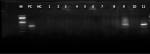
Detection of invA among isolates: all 11 suspected isolates that were recovered from preliminary tests were screened for the invA gene. Only the 2 isolates identified as Salmonella by Microbact also showed the expected invA bands. (Figure 1).
Two Salmonella isolates, which included 1(1.7%) in raw beef from Garki ward and 1(3.2%) in raw beef from Central area wards of Abuja were isolated in this study giving a prevalence of 1.5% for the 136 raw beef samples analyzed, none was isolated from 150 raw chicken samples. An overall prevalence of 0.7% of the 286 samples investigated is observed to be lower than recent studies [15-17] which reported prevalences of 11% in raw beef, 14.1% from commercial broiler chicken, and 15.4% in raw chicken respectively. Our investigation observed multiple drug resistance (MDR) in the two Salmonella isolates recovered. All the isolates (100%) were resistant to ampicillin, amoxicillin, cephalothin, sulphamethoxazole, tetracycline, trimethoprim and nitrofurantoin. One (1) of the isolates was resistant to tobramycin, gentamicin, colistin sulphate and cefotaxime. The resistance of Salmonella isolates to cephalosporins is emerging to be a significant public health problem, cephalosporins are considered to be one of classes of drugs of choice used in the treatment of invasive non-typhoidal Salmonella infections in situations where trimethoprim/sulfamethoxazole or ampicillin is clinically ineffective [23-25]. Research has shown that there is a linear correlation of resistance to beta-lactam antimicrobials with the lactamase level over some time and resistance to beta-lactam can be attained by escalating enzyme levels [26]. Hence, the protracted use or misuse of cephalosporins selects for resistance over a period. The MAR index reported in this study (0.45-0.5) seems to be high, which signals the exposure of the animal product source (cattle) to high-risk contamination with antibiotic-resistant pathogens from high-risk environments, which increases the possibility of shedding microorganisms harmful to humans [27].
The invasion gene invA was detected in both isolates. This gene is essential for full virulence in Salmonella and is thought to trigger internalization required for invasion of deeper tissue [23]. This finding closely agrees with other studies that reported the detection of this gene in almost all Salmonella isolates, [8,23], and reported that PCR assay using invA primers specific for Salmonella considerably decreases the number of false-positive results. Amplification of the invA gene is now recognized as an international standard procedure for the detection of the Salmonella genus [8]. Several studies in Nigeria have reported recovery of Salmonella spp. from apparently healthy animals and animal products at varying prevalence; 6.4% from chicken, 14.1% in raw meat, 39% in apparently healthy slaughtered food animals and 2% in raw beef respectively [11,13,28-29]. Other studies carried out in other parts of Africa have continued to report Salmonella from animals and animal products. Salmonella was detected with a prevalence of 19% in beef carcasses in South Africa [30], 4% in sausage, 2% in spiced meat minced meat in Egypt [31] and 4.2% in slaughtered cattle and 12.1% in minced meat in Ethiopia [32]. This varying prevalence may be attributed to the Salmonella carriage among animals in different locations and countries, and may be influenced by multiple aspects such as the critical role of the slaughter process in contamination of carcasses, abattoir environment, poor slaughter techniques, wet and dry hides and skin, contaminated slaughter equipment, improper evisceration, faulty transportation, and retail procedures, storage conditions, sample types, culture methods and culture media used[33].
The storage temperature of the samples collected in this study could be a factor that may have contributed to the low prevalence rate observed, which is an important factor in the survival of pathogenic bacteria [34]. The temperature range for the growth of Salmonella spp. is 5.2-46.2°C, with the optimal temperature being 35-43�C. Although freezing affects Salmonella spp. survival, it does not guarantee the destruction of the organism. There is an initial sharp decrease in the number of viable organisms at conditions close to the freezing point as a result of the freezing damage, but Salmonella spp. in some situations have been seen to have the ability to survive long term frozen storage at lower temperatures as well [35]. Another contributing factor may be linked to a limitation observed in this study; samples for this study were collected from registered fast-food restaurants and during sampling, it was discovered that some fast-food restaurants were not registered with AFFCON. It was also observed that the 2 Salmonella isolates identified by Microbact were confirmed to be Salmonella based on PCR detection of the inv A gene; this suggests that the Microbact is a sensitive tool for rapid identification from a pure culture. The need for rapid identification of organisms from samples with or without outbreaks is necessary, and thus the convenience and efficiency demonstrated by the Microbact kit suggests a possibility for it to be used for rapid identification of foodborne organisms and also can be used to improve diagnosis in laboratory settings.
Our study was able to provide new information on an important foodborne pathogen in the food industry, specifically fast-food restaurants, which were hitherto not reported in Abuja. The MDR status of the isolates and the presence of a virulence gene reported in this study is also significant. The inappropriate application and control of a uniform food safety management system like the Hazard Analysis Critical Control Points (HACCP) by some of the fast-food restaurants sampled and the subsequent recovery of a pathogenic organism from animal products used in fast-food restaurants is a cause for concern. Food animals are the major reservoirs of Salmonella and the findings in this study suggest that meat sourced and kept in an unhygienic environment, and kept in inappropriate storage conditions could be a potential reservoir for Salmonella [36]. The consumption of contaminated food of animal origin can bring about the acquisition of antimicrobial-resistant foodborne pathogens [37] which is undesirable. Antimicrobial use and misuse have been contemplated to be the most vital selecting force for antimicrobial resistance of bacteria development and spread in both veterinary and human medicine [38]. Inappropriate antibiotic usage in food animals could predispose humans to risks of antibiotic-resistant bacterial infections with the situation additionally complicated by the potential of resistant bacteria to transfer their resistance determinants to resident human microflora and other pathogenic bacteria [37]. It is, therefore, necessary for all fast-food restaurants in Nigeria to properly implement a food safety management system. Lack of personal hygienic measures, poor hygiene practices in abattoirs, lack of disinfection during retailing/storage equipment, poor handling and processing methods coupled with poor environmental sanitation are all potential factors that can lead to contamination of meat. Hence, proper handling of raw meat is encouraged to prevent Salmonella contamination. Further studies should be carried out to assess risk factors involved in the spread of MDR Salmonella isolates along the food chain.
Two (2) isolates of Salmonella were isolated from raw beef and all the isolates carried the virulence-associated gene invA and this suggests that they are potentially virulent. These isolates also exhibited multiple drug resistance (MDR) phenotypes. Overall it can be concluded that the prevalence of Salmonella in the Federal Capital Territory (FCT) seems to be lower than reported in recent studies.
What is known about this topic
- The main reservoirs of zoonotic Salmonella are food animals;
- The inappropriate use of antibiotics in animals has led to a threat of AMR in animal and human health.
What this study adds
- This study presents new data on the current prevalence of Salmonella from the meat used in fast-food restaurants in Abuja, Nigeria and contributes to the very scarce data on contamination of meat used for the preparation of meals in fast-food restaurants;
- The study shows a very high level of antibiotic resistance in Salmonella which should be further explored;
- This study confirmed the presence of invA virulence gene in the isolates which reinforces the need for the adoption of food safety management systems by all actors along the food supply chain, from farm to fork to limit the spread of such pathogens.
The authors declare no competing interests.
Salome Samuel Bawa and Jacob Kwada Paghi Kwaga contributed to the conceptualization, data curation, formal analysis, investigation, methodology, resources, and writing of the original draft. Jacob Kwada Paghi Kwaga and Mohammed Kabiru Lawan contributed to data curation, project administration, supervision, validation, writing - review and editing while Samuel Bitrus Bawa contributed to interpreting results and investigation. All authors have read and agreed to the final manuscript.
We are grateful to the Department of Public Health, Health, and Human Services Secretariat, Federal Capital Territory Administration, Garki Abuja for facilitating sample collection.
Table 1: distribution of fast-food restaurants and recovery frequency of samples
Figure 1: detection of Salmonella by PCR
- Heredia Norma, García Santos. Animals as sources of food-borne pathogens: a review. Animal Nutrition. 2018; 4(3):250-255.. PubMed | Google Scholar
- World Health Organization. WHO estimates of the global burden of foodborne diseases.WHO. Accessed 10 June 2020.
- Harakeh Steve, Yassine Hadi, Gharios Maya, Barbour Elie, Hajjar Shadi, El-Fadel Mutasem et al. Isolation, molecular characterization and antimicrobial resistance patterns of Salmonella and Escherichia coli isolates from meat-based fast food in Lebanon. Science of the Total Environment. 2005;341(1-3):33-44. PubMed | Google Scholar
- Plym Forshell, Wierup Martin. Salmonella contamination: a significant challenge to the global marketing of animal food products. OIE Revue Scientifique et Technique. 2006;25(2):541-554. PubMed | Google Scholar
- Desta Sesay, Mekonnen Addis. A review on major food borne bacterial Illnesses. Journal of Tropical Diseases. 2015;3(4):1-7.
- Angulo Frederick, Jones Timothy. Eating in Restaurants: a risk factor for foodborne disease. Clinical Infectious Diseases. 2006;43(10):1324-1328. PubMed | Google Scholar
- Njie Ateba Collins, Mochaiwa Biotumelo. Use of invA Gene specific PCR analysis for the detection of virulent Salmonella species in beef products in the North West Province, South Africa. Journal of Food and Nutrition Research. 2014;2(6):294-300. Google Scholar
- Amini Kumarss, Taghi Zahraei Salehi, Gholamreza Nikbakht, Reza Ranjbar, Javid Amini, Shahrnaz Banou et al. Molecular detection of invA and spv virulence genes in Salmonella enteritidis isolated from human and animals in Iran. African Journal of Microbiology Research. 2010;4(21):2202-2210. Google Scholar
- Olovo Chinasa, Reward Eleazar, Obi Scholastica, Ike Anthony. Isolation, identification and antibiogram of Salmonella from cloacal swabs of free range poultry in Nsukka, Nigeria. Journal of Advances in Microbiology. 2019;17(1):1-9. Google Scholar
- Adeyanju Gladys Taiwo, Ishola Olayinka. Salmonella and Escherichia coli contamination of poultry meat from a processing plant and retail markets in Ibadan, Oyo state, Nigeria. SpringerPlus. 2014;3(1):139. PubMed | Google Scholar
- Akeem Ahmed, Moshood Raji, Paul Mamman, Clara Kwanashie, Ibrahim Raufu, Abdulfatai Aremu et al. Salmonellosis: Serotypes, prevalence and multi-drug resistant profiles of Salmonella enterica in selected poultry farms, Kwara State, North Central Nigeria. Onderstepoort Journal of Veterinary Research. 2019;86(1):1667. PubMed | Google Scholar
- Raufu Ibrahim, Fashae Kayode, Ameh James, Ambali Abdulganiyu, Ogunsola Folashade, Coker Akitoye et al. Persistence of fluoroquinolone-resistant Salmonella enterica serovar Kentucky from poultry and poultry sources in Nigeria. Journal of Infection in Developing Countries. 2014; 8(3):384-388. PubMed | Google Scholar
- Olorunjuwon Bello, Temitope Bello, Oluwatosin Amolegbe, Shakirullah Sanwo. Bacteriological assessment of raw meats sold in Lagos, Nigeria. Scientia Agriculturae. 2016;16(1):20-25. Google Scholar
- Tafida Salome Yakubu, Kabir Junaidu, Kwaga Jacob Kwada Paghi, Bello Mohammed, Umoh Victoria Jarlarth, Yakubu Sabo et al. Occurrence of Salmonella in retail beef and related meat products in Zaria, Nigeria. Food Control. 2013;32:119-124. Google Scholar
- Bata Shalangwa Ishaku, Karshima Ngutor Solomon, Yohanna Jethro, Dashe Moses, Pam Victoria Adamu, Ogbu Kenneth Ikejiofor. Isolation and antibiotic sensitivity patterns of Salmonella species from raw beef and quail eggs from farms and retail outlets in Jos, Plateau State, Nigeria. Journal of Veterinary Medicine and Animal Health. 2016;8(4):29-34. Google Scholar
- Fagbamila Idowu Oluwabunmi, Barco Lisa, Mancin Marzia, Kwaga Jacob, Ngulukun Sati Samuel, Zavagnin Paola et al. Salmonella serovars and their distribution in Nigerian commercial chicken layer farms. PLOS ONE. 2017;12(3):e0173097. PubMed | Google Scholar
- Ogu Gideon, Akinnibosun Faith. Occurrence of Salmonella in Raw Chicken Meat from Retail Equipment and Environments in Southern Nigeria Open Markets. Notulae Scientia Biologicae. 2019; 11(2):175-182. Google Scholar
- Academia. Cowan and Steels Manual for the identification of medical bacteria. Accessed 10 March 2019.
- O´Hara Caroline. Manual and automated instrumentation for identification of Enterobacteriaceae and other aerobic gram-negative bacilli. Clinical Microbiology Reviews. 2005;18(1):147-162. PubMed | Google Scholar
- Clinical and Laboratory Standards Institute. Performance standards for antimicrobial susceptibility testing. 2017. Accessed 16 May 2020.
- Saba Riaz, Muhammad Faisal, Shahida Hasnain. Antibiotic susceptibility pattern and Multiple antibiotic resistances (MAR) calculation of extended spectrum ?lactamase (ESBL) producing Escherichia coli and Klebsiella species in Pakistan. African Journal of Biotechnology. 2011;10(33):6325-6331.
- Sweeney Michael, Lubbers Brain, Schwarz Stefan, Watts Jeffrey. Applying definitions for multidrug resistance, extensive drug resistance and pandrug resistance to clinically significant livestock and companion animal bacterial pathogens. Journal of Antimicrobial Chemotherapy. 2018; 73(6):1460-1463. PubMed | Google Scholar
- Rahn Kris, De Grandis Stephanie, Clarke Robert, McEwen Scott, Galán Jorge, Ginocchio Christine et al. Amplification of an invA gene sequence of Salmonella typhimurium by polymerase chain reaction as a specific method of detection of Salmonella. Molecular and Cellular Probes. 1992;6(4):271-279. PubMed | Google Scholar
- Calayag Alyzza Marie, Paclibare Phyllis Anne, Santos Pauline Diane, Bautista Corinne Aimee, Rivera Windell. Molecular characterization and antimicrobial resistance of Salmonella enterica from swine slaughtered in two different types of Philippine abattoir. Food Microbiology. 2017;65:51-56. PubMed | Google Scholar
- Van Thi Thu Hao, Nguyen Hoang Nam Kha, Smooker Peter, Coloe Peter. The antibiotic resistance characteristics of non-typhoidal Salmonella enterica isolated from food-producing animals, retail meat and humans in South East Asia. International Journal of Food Microbiology. 2012;154(3):98-106. PubMed | Google Scholar
- Hamilton Russell, Hulsebus Holly, Akbar Samina, Gray Jeffrey. Increased resistance to multiple antimicrobials and altered resistance gene expression in CMY-2-positive Salmonella enterica following a simulated patient treatment with ceftriaxone. Applied and Environmental Microbiology. 2012;78(22):8062-8066. PubMed | Google Scholar
- Sarina Pignato, Maria Anna Coniglio, Giuseppina Faro, Martine Lefevre, François-Xavier Weill, Giuseppe Giammanco. Molecular Epidemiology of Ampicillin Resistance in Salmonella spp. and Escherichia coli from Wastewater and Clinical Specimens. Foodborne Pathogens and Disease. 2010;7(8):945-51. PubMed | Google Scholar
- Jajere Saleh Mohammed, Adamu Nuhu Bala, Atsanda Naphtali Nayam, Onyilokwu Samson Amali, Gashua Muhammad Mamman, Hambali Idris Umar et al. Prevalence and antimicrobial resistance profiles of Salmonella isolates in apparently healthy slaughtered food animals at Maiduguri central abattoir, Nigeria. Asian Pacific Journal of Tropical Disease. 2015;5(12):996-1000. Google Scholar
- Adesiji Yemisi, Alli Oyebode, Adekanle Margaret, Jolayemi Justina. Prevalence of Arcobacter, Escherichia coli, Staphylococcus aureus and Salmonella species in Retail Raw Chicken, Pork, Beef and Goat meat in Osogbo, Nigeria. Sierra Leone j biomed res. 2011;3(1):8-12. Google Scholar
- Jaja Ishmael Festus, Bhembe Nolwazi Londiwe, Green Ezekiel, Oguttu James, Muchenje Voster. Molecular characterisation of antibiotic-resistant Salmonella enterica isolates recovered from meat in South Africa. Acta Tropica. 2019;190:129-136. PubMed | Google Scholar
- Abd El-Atty Nasser, Meshref Arafa Meshref Soliman. Prevalence of Salmonella and E.coli O157 in some foods. Journal of Veterinary Medical Research. 2008;18(1):73-78. Google Scholar
- Molla Bayleyegn, Alemayehu Daniel, Salah Woubit. Sources and distribution of Salmonella serotypes isolated from food animals, slaughterhouse personnel and retail meat products in Ethiopia: 1997-2002. Ethiopian Journal of Health Development. 2003;17(1):63-70. Google Scholar
- Wheatley Paul, Giotis Efstathios, McKevitt Aideen. Effects of slaughtering operations on carcass contamination in an Irish pork production plant. Irish veterinary journal. 2014 Jan 18;67(1):1. PubMed | Google Scholar
- Arrus Katia, Holley Richard, Ominski Kimberly, Tenuta Mario, Blank Gregory. Influence of temperature on Salmonella survival in hog manure slurry and seasonal temperature profiles in farm manure storage reservoirs. Livestock Science. 2006; 102(3):226-236. Google Scholar
- Oliver Stephen, Jayarao Bhushan, Almeida Raul. Foodborne pathogens in milk and the dairy farm environment: Food safety and public health implications. Foodborne Pathogens and Disease. 2005;2(2):115-129. PubMed | Google Scholar
- Zamora-Sanabria Rebeca, Alvarado Aandrea Molina. Preharvest Salmonella Risk Contamination and the Control Strategies: Current Topics in Salmonella and Salmonellosis. 2017. Google Scholar
- Mead Paul, Slutsker Laurence, Dietz Vance, McCaig Linda, Bresee Joseph, Shapiro Craig, Griffin Patricia, Tauxe Robert. Food-related illness and death in the United States. Emerging Infectious Diseases. 1999;5(5):607-625. PubMed | Google Scholar
- Okeke Iruka, Klugman Keith, Bhutta Zulfiqar, Duse Adriano, Jenkins Phillip, O´Brien Thomas et al. Antimicrobial resistance in developing countries. Part I: recent trends and current status. Lancet Infectious Diseases. 2005; 5(9):568-580. PubMed | Google Scholar
Search

This article authors
On Pubmed
On Google Scholar
Citation [Download]
Navigate this article
Similar articles in
Key words
Tables and figures
Article metrics
Recently from the PAMJ-OH